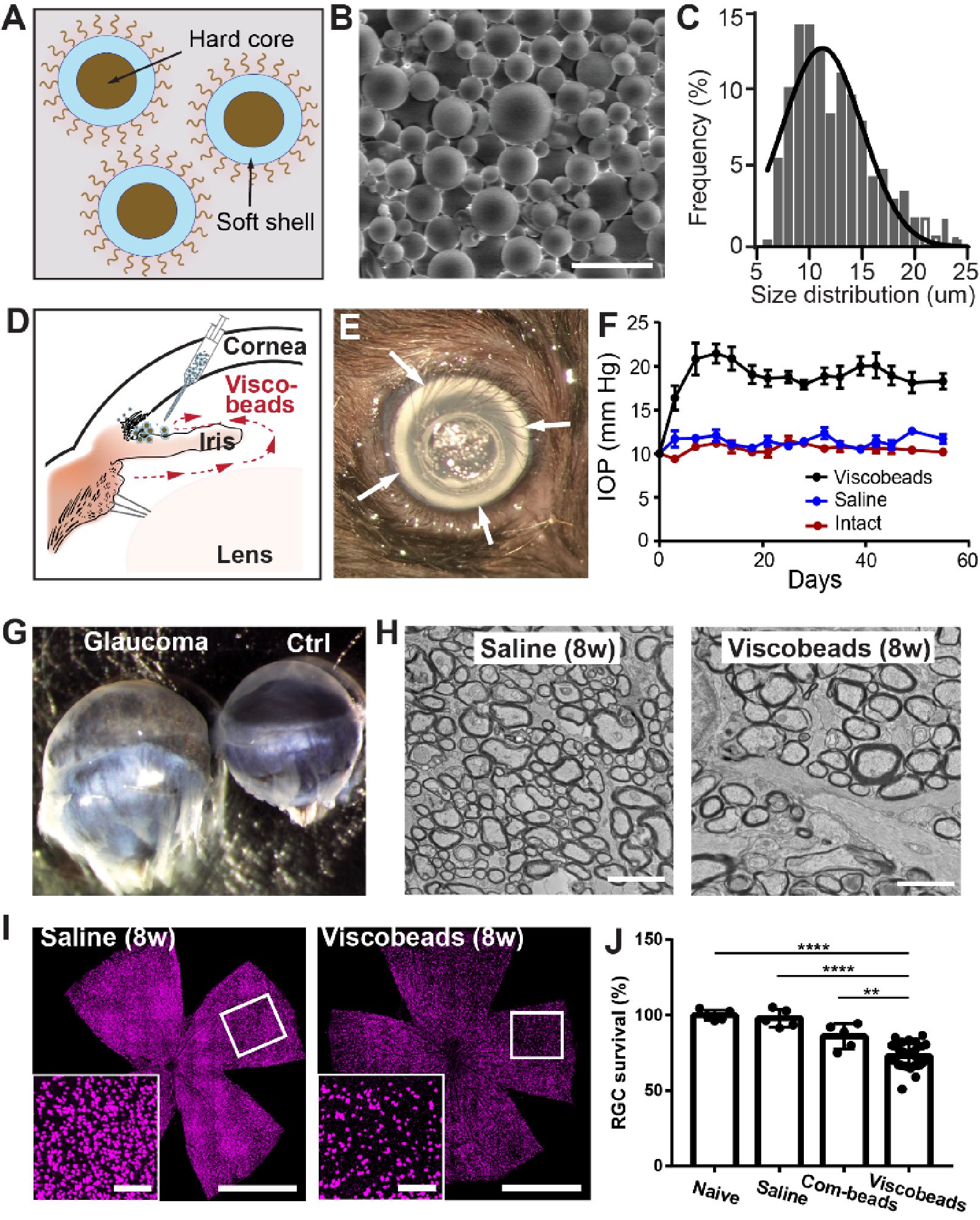
Injectable Viscobeads Technique for an Experimental Glaucoma Model

Current microbead occlusion models for glaucoma research exhibit significant variability and require multiple injections to maintain long-term ocular hypertension. The rigidity of polymeric beads and their mismatched size with trabecular meshwork (TM) structures often lead to insufficient stability of beads-tissue contact and unsatisfactory retention in TM tissues. To address these challenges, we have developed polymeric viscobeads with self-produced viscoelastic materials and a heterogeneous size distribution. This technique provides an accessible and versatile in vivo platform, facilitating the study of neurodegenerative mechanisms underlying glaucomatous progression and holding promise for future translational research applications. 

To request the viscobeads sample, please contact Dr. Wang at qbwang@binghamton.edu.

Non-Invasive Approaches for Early Detection of Neurodegeneration in Glaucoma

The identification of abnormal neural activities at an early stage holds significant potential for disease intervention and prevention. However, the current lack of adequate diagnostic methods hinders effective and timely intervention to protect against glaucomatous vision loss. In most cases, the early onset of symptoms remains undetectable until severe structural damage and vision loss manifest at advanced stages. To address this challenge, our research aims to establish a novel non-invasive electroretinography recording technique capable of identifying a comprehensive set of electrophysiological biomarkers. By elucidating both "high-risk markers" preceding retinal ganglion cell (RGC) loss and "functional loss markers" during glaucoma progression, we intend to obtain electrophysiological markers that enable early detection of glaucoma. This research has the potential to revolutionize glaucoma diagnostics, providing critical opportunities for early intervention and improved management of the disease.

Gene Delivery to the Eye for Glaucoma Treatment

The inner limiting membrane (ILM) acts as a physical barrier, limiting the access of therapeutic agents to target cells, thereby reducing the efficacy of gene therapy in treating retinal ganglion cell (RGC)-related retinal diseases such as glaucoma. Overcoming this challenge and enhancing ILM penetration in ocular gene delivery systems are crucial for the success of these therapies. In this research direction, our goal is to develop a noninvasive ultrasonic retinal stimulation system that improves the efficiency of ocular gene delivery into RGCs, offering potential applications for the treatment of glaucoma. Our approach involves utilizing a multiple-element 2D array transducer to create retina-shaped mechanical stimulation patterns, thereby enhancing the penetration of adeno-associated virus (AAV) vectors through the ILM. By addressing these key barriers, this research has the potential to advance gene therapy as a viable treatment option for glaucoma and other RGC-related retinal diseases.

Soft Bioelectronics for Intraspinal Neural Recording In Vivo

Our team develops bioinspired soft materials to address challenges in neural interfacing. In collaboration with partner groups, we engineered a polyvinyl alcohol (PVA) hydrogel–based optical neural probe and conductive hydrogel fibers by integrating carbon nanotubes into fatigue-resistant PVA matrices. When implanted in the lumbar ventral horn, these hydrogel microelectrodes enabled stable, long-term single-unit neural recordings in both anesthetized and freely moving animals. This work demonstrates the potential of hydrogel-based bioelectronics for chronic, high-fidelity neurophysiological monitoring in naturally behaving subjects.